Magnetic resonance
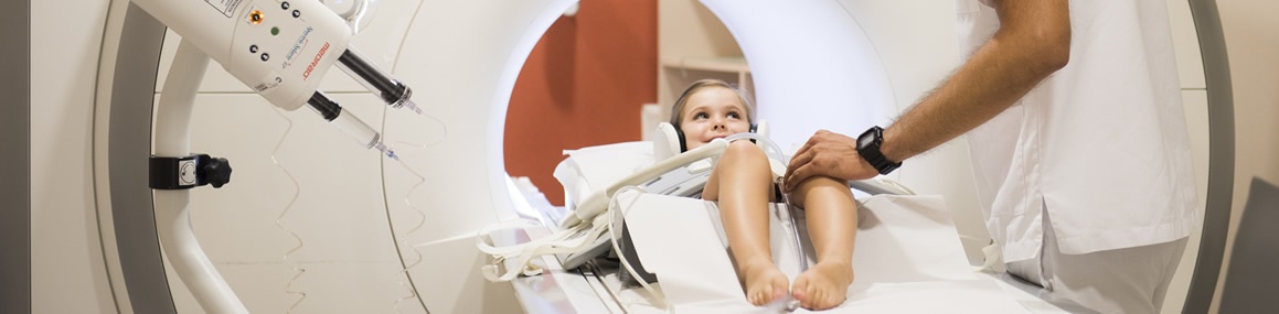
It is a painless diagnostic test that enables visualization of the interior of an area of the body (skull, thorax, abdomen, bones, etc.) using the magnetic resonance technique. This technique obtains the images by stimulating the organism via the action of an electromagnetic field produced by a powerful magnet and emission of radio waves. In some cases, to better delimit the structures of the body, it is necessary to administer a paramagnetic intravenous contrast.
How it is performed?
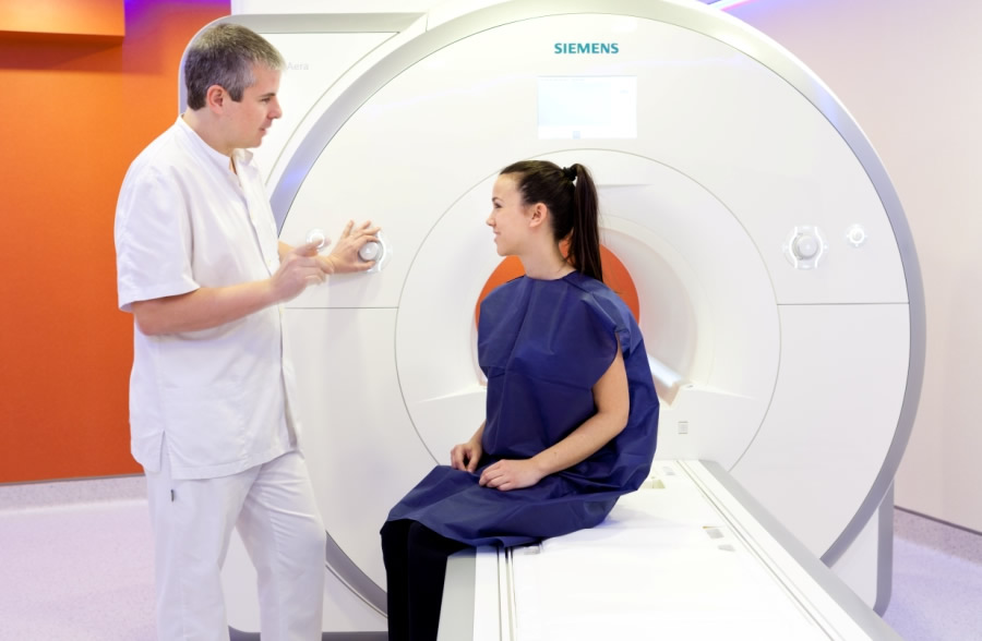
The patient lies down, face up on a gurney that is inserted into the magnetic resonance unit where the magnetic field is generated.
The scan usually lasts a total of about 30 minutes. In that time, the taking of images is not continuous but different image planes are made. The patient can communicate always with the technician performing the test.
If the examination requires contrast, the patient must not eat anything for two hours before the test.
What discomfort can one cause?
Magnetic resonance imaging is painless and is not invasive. Unlike other diagnostic techniques, it does not emit ionizing radiation.
The main inconvenience can come from the shape of the MRI tube, which may not be tolerable for people suffering from claustrophobia. If that is your case, please let the radiologist know, so they can look after you.
The equipment generates an intense noise, which is lessened with a pair of ear protectors.
What risks can one cause?
Magnetic resonance is contraindicated in people who are carriers of certain metal objects, especially pacemakers, some type of heart valves, cerebral ventricular leads or acupuncture needles.
If you are a carrier of any of these objects mentioned or of any other metallic object (prosthesis, nails, etc.), or if you have undergone any surgical intervention, please let the technician know.
The administration of contrast may, very rarely, cause a mild allergic reaction.
What alternatives are there?
There are other diagnostic imaging techniques (ultrasound, scanner, radiology, scintigraphy, etc.), each of which is indicated for a specific diagnostic orientation.
List of test
Heart
- Cardio-magnetic resonance
Interventional diagnosis
- Shoulder MR-arthrography
- Wrist MR-arthrography
- Hip MR-arthrography
Face and neck
- A.T.M. MR
- T.S.A. MR-angiography
- Vertebrae-cervical magnetic resonance
- Neck magnetic resonance
- Orbital magnetic resonance
- Dynamic T.S.A. MR-Angiography
Feminine pelvis
Abdomen
- Cholangiography MR
- Abdomen MR
- Abdominal MR-angiography
Cranium
- Intracranial MR-angiography
- MR cerebral spectroscopy
- MR Internal Auditory Canal (IAC)
- Cranial magnetic resonance
- Dynamic L.R.C. magnetic resonance
- Hypophysis magnetic resonance
- Orbital magnetic resonance
- Cerebral perfusion magnetic resonance
- MR of cerebral diffusion
- MR-angiography intracranial
Bones, joints and muscles
- Knee magnetic resonance
- Shoulder MR
- Forearm MR
- A.T.M. MR
- Arm MR
- Hip magnetic resonance
- Elbow MR
- Hand MR
- Wrist MR
- Foot magnetic resonance
- Leg MR
- Ankle magnetic resonance
- Thigh MR
- Shoulder MR-arthrography
- Wrist MR-arthrography
Spine
- Vertebrae-cervical magnetic resonance
- Dorsal vertebrae magnetic resonance
- Lumbar vertebrae magnetic resonance
Arteries and veins
- Abdominal MR-angiography
- Peripheral MR-angiography
- Thoracic MR-angiography
- Intracranial MR-angiography
- T.S.A. MR-angiography
Breast
- Mammary MR
Thorax
- Thoracic MR-angiography
- Thorax magnetic resonance